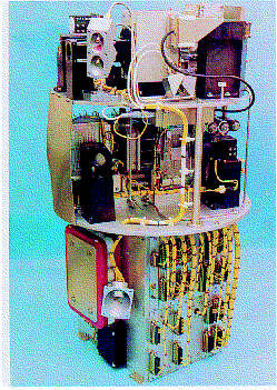
Fig. 3.1. Experiment Module TEM 06-5. (DASA)

Most of the nationally-funded German experiments were performed in one of the multipurpose experiment modules of the TEM 06 series. Each module consists of experiment platform, autonomous power supply and electronics package. The experiment-specific hardware is mounted on the platform, and is able to accept one or more sledges carrying biological samples as late as 30 min before launch. On request, the experiment-specific units can be manipulated during flight via telecommand. The different experiment set-ups of TEM are shown in Table 3.2. An identical ground model allows 1 g control experiments to be carried out in the launch site's laboratory.
Table 3.2. TEM experiment set-ups.
Field of Research                                     Experiment Module
--------------------------------------------------------------------------------------------

Free-flow electrophoresis                             TEM 04-1 (former 06-13) and TEM 04-2
Electro-cell-fusion                                   TEM 06-5 and TEM 06-11
Frog egg fertilisation and development                TEM 06-15
Microscopic observation of cells and injection of     TEM 06-5, TEM
06-16 and TEM 06-19
chemicals (markers, fixative) into samples
Biochemical investigation of cells in suspension      TEM 06-21
Chemical fixation of organisms (e.g. plant seedlings) TEM BIO/TEM KT
The TEM modules were financed by the German Ministry of Research and Technology, the German Space Agency DARA GmbH and ESA. The TEM KT and TEM BIO modules were manufactured by Kayser- Threde GmbH, Munich. All other TEM modules were manufactured by Daimler-Benz Aerospace AG, Space Infrastructure Division, Bremen (the former MBB-ERNO Raumfahrttechnik). Some representative modules are described below.
Experiment Module TEM 06-5
Length: 605-809 mm
Diameter: 403 mm
Mass: 47-56 kg
This module (Fig. 3.1)
was used primarily for fusion of plant protoplasts by the
electro-cell-fusion method. There are three independent
experiment units mounted on two platforms. Three sample cuvettes
are temperature-controlled before (6-8°C) and during
(20°C) flight. One cuvette can be observed via a microscope
(magnification up to 400x), with the transmitted images allowing
the experimenter to focus and select the best field of view by
telecommand. The cells can be stirred to avoid sedimentation
before the onset of µg. During the µg phase, the cells
are pumped from storage units into the fusion chambers to be
aligned in chains between the electrodes by an AC field. DC
pulses induce cell fusion.
Fig. 3.1. Experiment Module TEM 06-5. (DASA)
Experiment Module TEM 06-11
Length: 673 mm
Diameter: 403 mm
Mass: 54 kg
This module (Fig. 3.2)
was developed for the fusion of yeast and animal cells by
electro-cell-fusion, and for the transfection of genes into
cells. Three experiment units contain 15 fusion chambers where
the samples are temperature-controlled before and during flight
(22°C). The sample cuvettes are rotated to avoid
sedimentation of cells before the onset of µg. AC fields are
applied during µg to shepherd the cells into lines before
fusion by DC pulses.
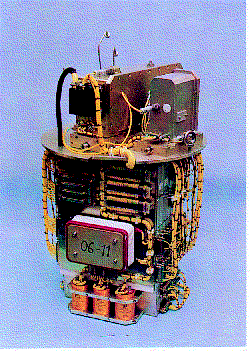
Fig. 3.2. Experiment Module TEM 06-11. (DASA)
Experiment Modules TEM 06-16 and 06-19
             TEM 06-16       TEM 06-19
 Length:     462 mm          522 mm
 Diameter:   403 mm          403 mm
 Mass:        34 kg           32 kg
These modules (Figs. 3.3/3.4) allow the microscopic observation of small organisms such as algae rhizoids or free- swimming cells. Some of the sample cuvettes can be supplied with chemicals, e.g. fixatives, during flight. A microscope (magnification up to 400x) observes the organisms using different illumination and contrast methods (e.g. phase contrast, darkfield). The realtime CCD camera image is transmitted to the ground, where the experimenter can focus and select the best field of view via telecommand. Up to five sample cuvettes (one for observation, four for e.g. fixation) can be housed in the module. All samples are temperature-controlled (e.g. 23°C). If necessary, sample cuvettes can be rotated before µg onset to avoid cell sedimentation. In addition to the microscope's illumination, further light sources can be used, e.g. for experiments investigating the interaction of gravitaxis and phototaxis of cells.
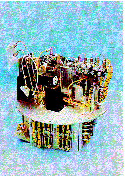
Fig. 3.3. Experiment Module TEM 06-16. (DASA)
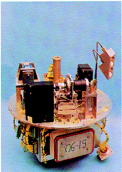
Fig. 3.4. Experiment Module TEM 06-19. (DASA)
Experiment Module TEM 04-1 (formerly 06-13)
Length:
1020 mm
Diameter: 403 mm
Mass: 87.5 kg
This module
(Fig. 3.5) was developed to investigate and optimise the process
of free-flow electrophoresis of cells. The main element is the
separation chamber, which requires precise isothermal conditions
(accuracy of 0.5°C throughout). The sample suspension and
buffer solution are transferred by different pumping systems. Two
CCD cameras observe the chamber's filling and separation
processes. The separation voltage is, for example, 1000 VDC at
100 mA and 600 VDC at 400 mA. The video records are analysed
post-flight to study µg's influence on the separation
process.
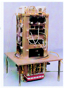
Fig. 3.5. Experiment Module TEM 04-1 (the former 06-13). (DASA)
Experiment Module TEM 06-21
Length: 470 mm
Diameter: 403 mm
Mass: 43 kg
Several dozen small
biological samples (e.g. suspensions of plant protoplasts) can
be processed during a rocket flight with this module (Fig. 3.6).
Changes in cell metabolism during various periods of µg
conditions can thus be investigated. A set of syringes is filled
with a cell suspension, while a second set contains chemicals
such as fixatives so that sample sets can be fixed inflight at
scheduled times. The samples are temperature-controlled before
and during flight.
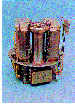
Fig. 3.6. Experiment Module TEM 06-21. (DASA)
Experiment Module TEM BIO
Length: 160 mm
Diameter: 403 mm
Mass: 22 kg
The chemical fixation of
organisms some centimetres long, such as plant seedlings, can be
performed with this module (Fig. 3.7). Four syringes filled with
fixatives are injected into four sample containers according to
a set time profile. The containers are temperature-controlled
before, during and after flight.
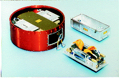
Fig. 3.7. Experiment Module TEM BIO. (DLR)
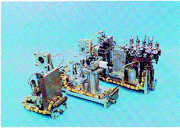
Fig. 3.8. Experiment Specific Insert for late access to the TEM 06-5 and TEM 06-19 Experiment Modules. (DASA)


 SP1206