Sea Urchin Eggs under Microgravity Conditions
H.-J. Marthy
Observatoire Oceanologique, CNRS (URA
2156) - UPMC, F-66650 Banyuls sur mer, France
Early events in sea urchin development were successfully
studied on three sounding rocket flights (MASER 4-6). The
principle aim was the identification of potential effects of
short exposure to microgravity on eggs at the fertilisation and
cleavage stages.
An initial experiment on MASER 4 in 1990 suggested that
fertilisation in µg would occur normally and that
morphological aberrancies in advanced larval stages (plutei with
strongly reduced arms) might be artifacts. The second experiment,
on MASER 5 in 1992, conclusively confirmed and completed these
results. There is no doubt that fertilisation sensu stricto does
occur correctly in a monospermic way in the absence of gravity.
Subsequent embryogenesis of such eggs on Earth leads to normal
pluteus larvae. Surprisingly, virgin eggs could not be fertilised
on the ground after exposure to µg.
The next experiment, on MASER 6 in 1993, studied whether
cleaving eggs (early embryos) are affected by short exposure to
µg. The results support the assumption that gravity changes
are effectively sensed by the individual embryonic cells, but
that development of the embryo as a whole is not affected. The
working hypothesis is postulated for future experiments that the
ability of embryonic cells in perceiving gravity is a cell cycle-
related, or even a cell cycle-dependent, phenomenon (interphase
or mitotic state). The experiments show that sounding rocket
flights provide suitable opportunities for studies in
developmental biology, with the proviso that precisely defined
developmental events are selected as subjects of research.
Introduction
Studies on fertilisation processes have so far been performed
aboard sounding rockets on two well known vertebrate and
invertebrate egg models: the mesolecithal amphibian and the
oligolecithal sea urchin egg. After the initial Spacelab D1
investigation¹ in 1985 into the fundamental question of
whether µg affects the fertilisation process in the clawed
toad Xenopus laevis, several sounding rocket experiments
from 1988 onwards gave conclusive results.2-8 The
author reports that fertilisation sensu stricto and the
establishment of the dorso-ventral polarity and of the bilateral
body symmetry are not directly affected by µg. However, a
causal relationship is likely to exist between µg exposure
of the eggs during fertilisation and a morphological phenomenon
at the gastrula stage (thickening of the epidermal blastocoelic
roof) and some deformities (distorted reduced tail) appearing at
the tadpole stage.
Thanks to flight opportunities offered by ESA, the substantial
support of CNES and multi-purpose space hardware developed for
the amphibian experiment,1, 9, 10 we were able to
approach similar questions beginning in 1990 with another classic
egg model: the sea urchin egg. In section 1, two experiments
related to the fertilisation process are covered. Section 2
considers potential µg effects on embryos at cleavage
stages.
1. FERTILISATION UNDER MICROGRAVITY CONDITIONS AND SUBSEQUENT
DEVELOPMENT ON THE GROUND (MASER 4-5)
Major principles of fertilisation in animal organisms,
including mammals, have been identified by studies on sea urchin
eggs.11-15 It was therefore a logical step to study
its monospermic fertilisation under µg conditions. Any new
result or observation from space experiments could be interpreted
against a whole range of previous biological, structural,
biochemical and molecular studies (for example, refs. 16-18). The
eggs of the species Paracentrotus lividus (ripe in spring
and in autumn) and Sphaerechinus granularis (ripe the
whole year) appeared particularly well suited.
Materials and methods
Biological material
15 (MASER 4) and 30
(MASER 5) adult sea urchins (Paracentrotus lividus) were
brought from the western Mediterranean Sea to ESRANGE. The
animals were placed either individually or in small groups in
plastic boxes filled with natural sea water. For transportation,
several boxes were placed together in isotherm containers at
about 16°C. The males and females were maintained separately
in two aquaria at ESRANGE below 16°C to avoid accidental
spawning.
Eggs and sperm were obtained by injecting the animals with
either 0.2 ml of 0.1 M solution of acetylcholine chloride and sea
water or by gently shaking. Sperm and eggs were released within
about 2 min (Figs. 1-1 & 1-2) and collected separately in Petri
dishes filled with filtered sea water. The Automatic Experiment
Containers (AECs) for fertilisation studies 10 were
loaded with pipettes from these dishes.

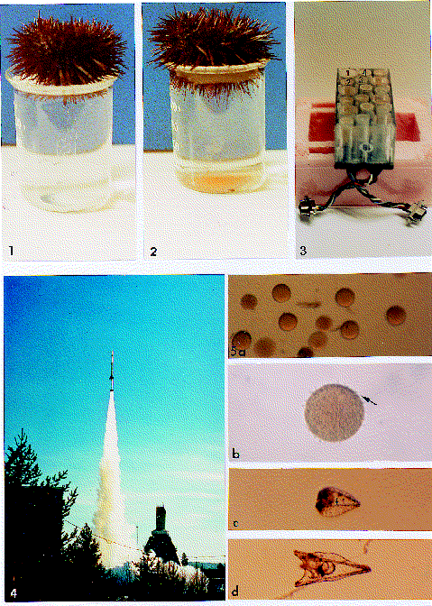
Fig. 1. The MASER 4 mission. Eggs and sperm are released (1-1
& 1-2) by adult sea urchins of the species Paracentrotus lividus
after injection of a solution of acetylcholine/sea water. Eggs
and sperm are separately stored and transferred to the various
chambers of the experiment block shown in Fig. 1-3. The Automated
Experiment Container (AEC), contained four identical
fertilisation units. In activating the plunger of chamber 3, sea
water was pushed, via the sperm chamber (2) towards the egg
chamber (1), where the eggs were inseminated. In activating the
plunger of chamber 4, a fixation solution was added to the egg
chamber. Fig. 1-4. Launch of MASER 4 on 29 March 1992 at 10.07
local time. (Photograph reproduced with kind permission of P.
Holm, Swedish Space Corporation.). Fig. 1-5. Basic post-flight
observations on the eggs fertilised in µg. 5a: flown virgin
eggs recovered live, no longer fertilisable. 5b: flown egg,
fertilised and fixed during the µg phase; arrow indicates
sperm attached to the fertilisation envelope. 5c: aberrant
pluteus larva, differentiated on ground, from an egg fertilised
in µg. The reduced arm growth is the most prominent defect;
however, the larval body with a skeleton has been formed (arrow).
5d: young pluteus differentiated from a ground control egg, at
day 7.
Experimental hardware and procedures
The
AEC used for the MASER 4 experiment was a modification of the
former multi-purpose space hardware developed by CCM (Nuenen, The
Netherlands) for the amphibian experiment.9 This
hardware (Fig. 1-3) was not totally satisfactory because of some
toxicity in the latex membranes of the culture chambers. However,
MASER 5's AEC (Fig. 2), specifically conceived and developed for
sea urchin eggs,19 yielded excellent results.


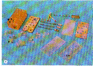
Fig. 2. Automated Experiment Container (AEC). Fig. 2-1.
Disassembled AEC hardware used on MASER 5. Photograph from P.
Gerrits, Bergeyk, NL, reproduced with kind permission of CCM,
Nuenen, NL. bl: experiment block. A plastic bag unit, filled with
sea water (s), sperm (arrow) and a fixative solution (f), is
inserted into the holes (s, e, f and arrow). Once the plastic
covers (c) are placed on the six plastic bag units, the cover
plate (co) and the mechanical-electronic plate (m) are attached.
The plungers (p) are mounted on plate m. Unmarked pieces are used
during the preparation procedures. The hardware is then
integrated into the CIS boxes.10 Fig. 2-2. Assembled
AEC flight hardware.
Loading the AECs was done 7 hr (MASER 4) and 10 hr (MASER 5)
before launch. Insemination of the eggs on MASER 4 was performed
automatically after 60 s of µg, followed by fixation of
parts of the egg samples 60 s before the end of the µg
phase, leaving 5 min of µg for the fertilisation process.
On MASER 5, insemination occurred after 60, 300 & 360 s during
the total of 420 s of µg, and the fixation plungers were
activated for parts of the samples 30 s before the end of
µg. For MASER 4-5, half of the egg samples fertilised during
µg were recovered alive. In both cases, samples of virgin
eggs and unused sperm were also recovered live.
Analysis
Initial post-flight examination
of the eggs was possible 3.5 hr after lift-off (Figs. 1-4 & 3-1).
Live recovered eggs were transferred to Petri dishes holding 5
ml of sea water and further development was recorded over 10 days
(MASER 4) and 42 days (MASER 5). Fixed material was stored for
structural analyses by Scanning and Transmission Electron
Microscopy (SEM and TEM).
Results and discussion
Pre-flight tests
It was found20
before the experiment that virgin eggs could best be stored at
5-11°C. The fertilisation rate was still high after about
12 hr, but it decreased rapidly after 24 hr. Dry' sperm stored
at 5°C could be used for at least 48 hr. In a recent
preservation study,21 we were able to prolong the
storage time for virgin eggs and dry sperm at 4°C for more
than 4 days.
Fertilised eggs exposed to 18 g hypergravity still showed
essentially normal embryonic and larval development. Vibration
tests indicated no harm to either virgin or fertilised eggs. As
parthenogenesis was not triggered by vibration, it could be
assumed that any effects on egg fertilisation and subsequent
embryogenesis should be interpreted as a result of µg.
MASER 4 experiment (Fig. 1)
Based on
elevation of the fertilisation envelope as the main gross
morphological criterion for a successful fertilisation, only a
low fertilisation rate of about 30% of the ground control samples
and of 0-10% of the flight samples was found. The causes were a
slight toxicity and delivery of the hardware only just before the
campaign began.
These low percentages made a final conclusion difficult.
However, SEM and TEM images (showing the presence of
fertilisation envelopes, extruded cortical granules and elongated
egg surface microvilli)22, showed that fertilisation
under µg most likely happened in the normal monospermic way,
triggering the whole cascade of fertilisation events (e.g. blocks
to polyspermy, initiation of embryonic development). Several egg
samples recovered live developed up to a pluteus stage, but
showed a strongly reduced arm growth (Fig. 1-5c).23
MASER 5 experiment (Figs.2 & 3)
The aim
of this experiment was to confirm and complete the results from
MASER 4 and, in addition, to identify possible µg effects
on intact spermatozoa and virgin eggs. The hardware was
completely redesigned for this purpose.19
The experiment was a complete success in both technical and
scientific terms. The AEC worked nominally in space and on the
ground at a constant temperature of 17°C inside the Cells
in Space boxes.10 Analysis of live egg samples
revealed a 95% fertilisation rate (Fig. 3.2-3.8), although the
elevation of the fertilisation membrane was occasionally weak.
The cleaving eggs continued embryonic and larval development.
Young pluteus larvae were swimming after 4 days, identical to the
controls. Larvae lifetimes could be increased to >40 days by
feeding with microalgae. Interestingly, flown virgin eggs could
no longer be fertilised. We have not pursued it being a µg
effect. Flown sperm maintained its fertilisation ability on fresh
virgin eggs. From the fixed samples, SEM and TEM studies
confirmed successful insemination and fertilisation.22

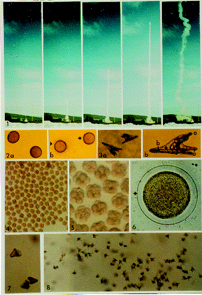
Fig. 3. The MASER 5 mission. Fig. 3-1. Launch of MASER 5 on
9 April 1992 at 12.07 local time. 317 km apogee, 7 min µg
phase. Recovery of CIS-3 experiment boxes and payload occurred
about 3.5 hr after launch. Fig. 3-2. Normal aspect of live eggs
in translucent light: 2a. virgin eggs; 2b. left, inseminated egg
surrounded by sperm (arrowhead); right, clearly fertilised eggs
showing a fertilisation envelope (arrow). Diameter 80 µm.
Fig. 3-3. Normal aspect of larvae at the pluteus stage, observed
in translucent light. 3a: two swimming fully differentiated
plutei at day 10. 3b: pluteus larva (a: anal arm with anal rod
inside. b: body rod, or calcite spicule (arrow). e: oesophagus.
i: intestine. o: oral lobe. s: stomach). Fig. 3-4. Immediate
post-flight aspect of fertilised egg samples, recovered live. The
eggs are at the cleavage stages. In total, about 40 000 eggs,
fertilised in space, were recovered alive and continued
development on the ground. An identical number served as ground
control. All were from the same female and fertilised with the
sperm of one male. Fig. 3-5. Eggs from another flown sample, at
8-16 cell stages of cleavage. No difference is visible between
the various samples. Fig. 3-6. Egg fertilised (inseminated) after
60 s in µg and fixed after 5.5 min. A well-elevated
fertilisation envelope surrounds the egg (arrow), formed by
exocytosis of cortical granules within 60 s of the sperm-egg
contact. It prevents polyspermy; the process is also known as
slow block.' The presence of a fertilisation envelope is a clear
indication of successful fertilisation. Fig. 3-7. Swimming larvae
at the prism/early pluteus stage (about 40 hr old),
differentiated on the ground from flown eggs. Fig. 3-8. Numerous
normal young plutei at day 11, developed on the ground from flown
eggs (compare with Fig. 3-3a/b). The developing larvae of flown
eggs and of ground controls were cultured in small Petri dishes.
From day 13 onwards, unicellular algae were offered as food. The
last flown-egg pluteus, which showed no sign of metamorphosis,
was lost at day 42.
Conclusion
Taking into careful account all observations on live and fixed
eggs/larvae, some firm conclusions can be drawn:
-
In sea urchin eggs, fertilisation under real µg conditions
occurs in a normal monospermic way and with a high (normal)
fertilisation rate.
- Eggs fertilised in µg develop
normally on the ground to an advanced pluteus stage.
- The
unspectacular results have to be interpreted in a positive way.
It is reassuring to know that a classic animal egg model clearly
shows that the initial crucial processes for creating new life
are not disturbed in µg.
- Despite the brief duration
of µg, sounding rocket flights are well suited to
developmental biology studies, with the proviso that clearly
defined processes are studied (e.g. fertilisation sensu
stricto, a specific developmental step such as egg cleaving,
and onset of gastrulation).
- It is an open question
whether the well known and considerable regulation capacities of
the sea urchin egg mask or compensate for some temporary µg
effects at fertilisation during subsequent embryonic and larval
development.
- It is also an open question whether eggs fertilised and
kept under continued µg conditions would be able to develop
normally. New space flight opportunities are necessary and
therefore solicited.
2. GROUND-BASED DEVELOPMENT OF SEA URCHIN EGGS AFTER EXPOSURE
TO MICROGRAVITY DURING CLEAVAGE STAGES (MASER 6)
Introduction
By selecting a precise portion of the development under study,
the few minutes of µg available during a sounding rocket
flight may serve for determining the potential effects of
weightlessness and thus for determining the role of gravity on
that particular developmental event.24 Bearing in mind
the step-by-step analysis of sea urchin development, the exposure
of cleaving eggs to µg appeared not only worthwhile as the
next step after the fertilisation study but also in the more
general context of how cells might perceive gravity forces. In
fact, an original hypothesis requires that the cell nucleus,
anchored in the cytoplasm, mechanically interferes with the
cytoskeleton and the cell membrane.25, 26 The
slightest deviation in the cytoskeletal organisation by the
nucleus, due to intracellular tension or pressure changes, would
lead firstly to possibly irreversible modifications of the
cytoskeleton and, ultimately, when the effect/s are amplified
throughout succesive cell generations (as occurs in
embryogenesis, for example), to severe morphogenetic
disturbances. Yet a nucleus-cytoskeleton-membrane inter-
relationship alone could hardly be the complete gravi-sensitive
system.27 The morphological situation had already been
revealed to be more complex because the nucleus as a compact mass
is present only during the interphase of the cell cycle but
absent throughout mitosis. The question was therefore whether
only interphase cells, and not mitotic cells, might be capable
of perceiving gravity. In practice, the aim of the study was to
check whether exposure of the cleaving eggs to a µg
environment leads to a disturbed morphogenesis, and whether a
disturbed morphogenesis results only when mg acts on interphase
cells. The large translucent eggs in early, synchronously
dividing, and in later, asynchronously dividing, cleavage stages
appeared well suited to this purpose.
Materials and methods
For the MASER 6 flight (Fig. 4), cleaving eggs of the
Sphaerechinus granularis sea urchin species were used.
Artificially fertilised on the ground at 11°C, intact
embryos at early and later cleavage stages were placed in the
hardware developed by CCM (Figs. 4-3 to 4-5; ref. 19). Besides
a passive static maintenance of parts of the samples, others were
placed on a 1 g centrifuge. A sample of eggs in early cleavage
was prepared for flight and ground video recording; the flight
recording failed for technical reasons. Identical samples were
prepared for ground controls. The total exposure to µg was
6 min. Parts of the samples were automatically fixed close to the
end of the µg phase; others were recovered live and kept in
culture for many weeks.

Fig. 4. The MASER 6 mission. Fig. 4-1. Launched on 4 November
1993 at 08.07 local time. 244 km apogee; 6 min µg phase. The
CIS-4 experiment boxes were recovered after 1.5 hr. Fig. 4-2.
Normal fully grown pluteus larvae of the sea urchin species
Sphaerechinus granularis from eggs exposed to µg at the
cleavage stages. Fig. 4-3. Two AECs with six culture chambers for
a passive maintenance of about 1000 eggs/chamber. Fig. 4-4.
Preparation of the 1 g onboard centrifuge (c) before insertion
into the experiment CIS-box (e). Fig. 4-5. Assembled hardware for
videorecording the cleaving eggs, before insertion into the CIS-
box. Fig. 4-6. Post-flight MASER 6 with its integrated CIS-4
segment. Fig. 4-7. The first post-flight view of living eggs at
their cleavage stages. These eggs developed into normal plutei
larvae, as shown in Fig. 4-2.
Analysis
Post-flight analyses focused on the continued development in
culture of the samples recovered live and on the quantitative
estimation of the egg samples fixed at the end of the µg
phase. In the first case it was expected that egg samples of
early cleavage stages, after exposure to the few minutes of real
µg, would develop into:
- 100% normal larvae, meaning that there was either no effect
or that all cells were only in mitosis, or
- 100% abnormal
larvae, meaning that there was either an effect on all cells or
that all cells were simultaneously in interphase, or
- into
normal or abnormal larvae, meaning that there was an effect in
one case (= interphase cells?) and none in the other (= mitotic
cells?). From egg samples of late cleavage stages, it was
expected that 100% should develop into abnormal larvae
(simultaneous presence of interphase and mitotic cells).
Results and discussion
Whatever the cleavage stage exposed to µg, all eggs
recovered live continued embryonic development and a high
percentage (>75%) differentiated into normal plutei (Fig. 4-
2). The plutei were fed from day 12 with the microalga
Isochrysis galbana (starvation occurs within about 3 weeks
without feeding). The last larvae died after 2.5 months of
culture, showing the first signs of metamorphosis.
How to interprete this clear result? Either there is no gravi-
sensitive nucleus-cytoskeleton-membrane relationship conceivable
on a mechanical basis, or there is such a relationship but the
effects of brief gravity changes are regulated and development
continues normally after return to normal gravity conditions. The
quantitative exploration of the fixed material gave a clue that
µg effects were produced (Fig. 5). In early cleavage samples
from the onboard 1 g centrifuge (FCE), no advancement of the
cleaving process was observed compared to the ground controls
(GFE). There was a slight tendency in 0 g samples (FFE) towards
more rapid cell division when compared to the GFE. It was clearer
when compared to the FCE. Comparing later stages (asynchronous
cell divisions), the 0 g samples (FFL) appear slightly advanced
compared to those of the 1 g onboard centrifuge (FCL) and the
ground control (GFL).
Fig. 5. Exposure to different g-levels (1 g ground, 0 g
flight, 1 g centrifuge) of sea urchin embryos at early and late
cleavage stages. Fixed early cleavage stages in 1 g (FCE),
compared to ground control (GFE) and 0 g (FFE) samples, show no
advancement of the cleavage process. A slight tendency for
acceleration appears in 0 g samples compared to the ground
controls. As to embryos at late cleavage stages, the samples in
0 g (FFL) appear to have a slight tendency for acceleration of
the cleavage process, compared to the ground control (GFL) and
the 1 g samples (FCL). Abbreviations NF: unfertilised eggs. F:
fertilised eggs. 2C: 2-cell stage. 4C: 4-cell stage. 8C: 8-cell
stage. MS: morulas. The evaluation considers only eggs recovered
intact.
Conclusion
One main conclusion is evident from observations on the
developing embryos and larvae: development of eggs exposed at
cleavage stages to short µg conditions proceeds normally.
Whether cleaving embryos in the interphase state or in mitosis
were exposed, no long-term effects appeared during the subsequent
embryonic and larval development on the ground. There were either
never any effects or they became regulated throughout
embryogenesis. Taking into account the quantitative results, this
latter assumption has to be favoured. Whereas the embryo as a
whole developed normally, the individual embryonic cells sensed
apparent alterations in gravity and responded by shortening (1
g centrifuge) or lengthening (0 g) the cell cycle or a phase of
it. Whether the interphase or the mitotic phase in embryonic
cells will be revealed as the gravi-sensitive state is a
challenging question for the future. The working hypothesis is
postulated that the ability of embryonic cells (and possibly of
other cell systems as well28) for perceiving gravity
is a cell cycle-related, or even a cell cycle-dependent,
phenomenon.
Acknowledgement
Thanks are due to ESA for the flight opportunities and to CNES
for substantial support. Particular mention must be made of
Project Manager Mr W. Herfs at ESA/ESTEC and of his assistant Dr
R. Demets (MASER 4 mission), as well of the mission scientists
Mr J. Vreeburg (NLR Amsterdam, NL) and Prof G. Frohberg (TU
Berlin, D). Various help is kindly acknowledged from Mr R.
Huijser and Mr L. van den Bergh (Fokker Space, Amsterdam, NL),
Mr H. Willemsen (CCM, Nuenen, NL) and his colleagues, as well as
from Mr J. Zaar and S. Anflo, Project Managers of the Swedish
Space Corporation. Last, but not least, many thanks to my team
members P. Schatt and U. Marthy (Banyuls-sur-mer, F), who were
directly involved in these projects.
References
-
Ubbels, G. A. (1988). The role of gravity in the establishment
of the dorso-ventral axis in the amphibian embryo. In Biorack
on Spacelab D1. 147-155. ESA SP-1091, ESA Publications
Division, Noordwijk, The Netherlands.
-
Ubbels, G. A. (1988). A first fertilisation of frog eggs in
space. Microgravity News from ESA, 1/2, 19-23.
-
Ubbels, G. A., Berendsen, W. & Narraway, J. M. (1989).
Fertilisation of frog eggs on a sounding rocket in space. Adv.
Space Res. 9 (11), 187-197.
-
Ubbels, G. A., Berendsen, W., Kerkvliet, S. & Narraway, J.
(1990). The first seven minutes of a Xenopus egg fertilized on
a sounding rocket in space. In Microgravity as a tool in
developmental biology. 228-230. ESA SP-1123, ESA Publications
Division, Noordwijk, The Netherlands.
-
Ubbels, G. A., Berendsen, W., Kerkvliet, S. & Narraway, J.
(1990). Fertilization of Xenopus eggs in space. In Life
Sciences Research in Space. 249-254. ESA SP-307, ESA
Publications Division, Noordwijk, The Netherlands.
-
Ubbels, G. A., Berendsen, W., Kerkvliet, S. C. J. & Narraway, J.
M. (1991). Fertilisation and development of eggs of the South
African clawed toad, Xenopus laevis, on sounding rockets
in space. Adv. Space Res. 12 (1), 181-195.
-
Ubbels, G. A., Reijnen, M., Meijerink, J. & Narraway, J. M.
(1994). Xenopus laevis embryos can establish their spatial
bilateral symmetrical body pattern without gravity. Adv. Space
Res., 14 (8), 257-269.
-
Ubbbels, G. A., Reijnen, M., Meijerink, J. & Narraway, J. (1995).
The role of Gravity in the Establishment of the dorso-ventral
axis in the Amphibian Embryo. In Biorack on Spacelab IML-
1. 175-185. ESA SP-1162, ESA Publications Division,
Noordwijk, The Netherlands.
-
Ubbels, G. A. & Willemsen, H. P. (1990). Automatic experiment
container for fertilization of frog eggs in space. A multi-
purpose piece of classified space hardware. In Microgravity
as a tool in developmental biology. 75-78. ESA SP-1123, ESA
Publications Division, Noordwijk, The Netherlands.
-
Huijser, R. L., Aartman, L. H. & Willemsen, H. (1990). Cells
in Space: Sounding Rocket Facilities for Cell Biology and
Biotechnology in Micro-gravity, 455-466, ESA SP-307, ESA
Publications Division, Noordwijk, The Netherlands.
-
Hsrstadius, S. (1973). Experimental Embryology of
Echinoderms. Clarendon Press.
-
Czihak, G. (1975). The sea urchin embryo. In Biochemistry and
Morphogenesis. Springer Verlag, Berlin.
-
Okazaki, K. (1975). Normal Development to Metamorphosis. In
The sea urchin embryo (ed. Czihak, G.). 177-266. Springer
Verlag, Berlin.
-
Giudice, G. (1986). The sea urchin embryo. A developmental
biological system. Springer Verlag, Berlin.
-
Wilt, F. H. (1987). Determination and morphogenesis in the sea
urchin embryo. Development 100, 559-575.
-
Alberts, B. (1989). Germ cells and fertilization. In Molecular
Biology of the Cell (2nd ed.). 839-878. Garland Publishing
Inc., New York and London.
-
Epel, D. (1990). The initiation of development at fertilization
(review). Cell Diff. and Dev. 29, 1-12.
-
Schatten, H. & Schatten, G. (eds.). (1989). The Cell Biology
of Fertilization (ISBN 0-12-622590-7) and The Molecular
Biology of Fertilization (ISBN 0-12-622595-8). In Cell
Biology: A series of Monographs. Academic Press.
-
Willemsen, H. (1994). Automatic Hardware for Biological
Experiments in Microgravity. ESA SP-366, 461-465.
-
Marthy, H.-J. (1989). On fertilization and development of sea
urchin eggs: a) in a space flight proved system and b) under
hypergravity conditions. ELGRA News 11, 79.
-
Anthony, P., Ausseil, J., Bechler, B., Bengurä, A.,
Blackhall, N., Briarty, L. G., Cogoli, A., Davey, M. R., Garesse,
R., Hager, R., Loddenkemper, R., Marchant, R., Marco, R., Marthy,
H.-J., Perry, M., Power, J. B., Schiller, P., Uglade, C.,
Volkmann, D. & Wardrop, J. (1996). Preservation of viable
biological samples for experiments in space laboratories. J.
of Biotechnol., 47 (2/3), 377-393 (authors in alphabetical
order).
-
Marthy, H.-J., Schatt, P. & Santella, L. (1994). Fertilization
of sea urchin eggs in space and subsequent development under
normal conditions. Adv. Space Res.14 (8), 197-208.
-
Marthy, H.-J. (1990). Microgravity and development of aquatic
animals. In Life Sciences Research in Space. 255-257. ESA
SP-307, ESA Publications Division, Noordwijk, The Netherlands.
-
Marthy, H.-J., Schatt, P. & Marthy, U. (1994). Sounding Rocket
Flights, suitable opportunities for development biology studies
in space. 193-196. ESA SP-355, ESA Publications Division,
Noordwijk, The Netherlands.
-
Mesland, D. (1990). Gravity Effects on Cells. In Life Sciences
Research in Space. 221-225. ESA-SP 307, ESA Publications
Division, Noordwijk, The Netherlands.
-
Bornens, M. (1991). Cytosquelette et Dynamique Cellulaire;
Biologie Gravitationnelle. In Rapport du Seminaire de
Prospective Microgravite du CNES, Aix-en-Provence, France.
-
Marthy, H.-J., Schatt, P. & Marthy, U. (1995). Development of sea
urchin eggs after exposure to microgravity during cleavage
stages. ELGRA News 19, 77.
-
Moore, D. & Cogoli, A. (1996). Gravitational and space biology.
Chapter 1 in Biological and Medical Research in Space. An
overview of Life Sciences Research in Microgravity (eds.
Moore, D., Bie, P. & Oser, H.). ISBN 3-540-60636-X, Springer
Verlag, Berlin.
About|
Search|
Feedback


 SP1206
Published April 1997.
Developed by ESA-ESRIN ID/D.